Tripping the light fantastic
Meet the unsung heroes of Imperial's
microscopy facility – making research visible.

Step into the ordinary looking, glass-fronted modern building just around the corner from the main entrance of the Science Museum and you could be in any workplace in the city. Ranks of wooden desks are populated by workers staring intently at their computer screens. Dotted around the desks are pictures of loved ones, a pair of once-used trainers, the full life and death cycle of a number of houseplants and, more incongruously, a red tricorn hat and yellow flower garland, perhaps from some vaguely remembered office party.
But the name above the main entrance – Sir Alexander Fleming Building – hints that this is no ordinary office. In fact, groundbreaking and potentially Nobel Prize-winning research takes place within its walls, because this is the Facility for Imaging by Light Microscopy (FILM), offering a suite of 15 state-of-the-art microscopes to support breakthrough research across Imperial. Last year, FILM microscopes were used for more than 11,000 hours by 400 scientists and other researchers from 160 different groups around Imperial. And the scope of its work is breathtaking.
“We can be working across five or six different areas every day,” says Dr David Gaboriau, a microscopy specialist at FILM. “In the morning we might image malaria parasites, heart cells and cancer treatments, and then in the afternoon, take movies of live neurons and other cell types in brain sections and nanoparticles inside cells. It is incredible to be on the cutting edge of such world-leading science every day. Sitting alongside Imperial’s scientists as they make their innovative discoveries is like seeing a work of art for the first time.”


Microscopic imagery

Human fibroblast cells, stained for mitochondria (magenta), microtubules (orange) and actin (green), imaged on a confocal microscope.

Human fibroblast cells, stained for mitochondria (cyan), microtubules (green) and actin (cherry), imaged by three dimensional structured illumination microscopy.

Microglia cells in a mouse brain section, imaged on a confocal microscope. Sample: Dr Mathieu Nollet.
The FILM team is composed of Professor Cristina Lo Celso and Professor Vania Braga, heads of the facility (Cristina is pictured right), Dr Volodymyr Nechyporuk-Zloy, facility manager, and two microscopy specialists, Gaboriau and Dr Ana Ferreira Da Silva. And the list of people and departments the Facility has worked with reads like a who’s who at Imperial. Many projects are being developed with the National Heart and Lung Institute, such as Dr Charlotte Dean’s work with confocal microscopy of lung slices; Dr Jorge Bernardino de la Serna’s work on understanding cell-cell interactions at the molecular levels, which promises better drugs to treat diseases like pneumonia and chronic obstructive pulmonary disease; and other collaborations on diseases such as idiopathic pulmonary fibrosis and asthma.
“When we capture something never seen before we’re helping prove a hypothesis or confirm that a paradigm is wrong,” says Lo Celso. “When I first visualised blood stem cells in the bone marrow of anaesthetised mice, for example, the working hypothesis was that they sat adjacent to osteoblasts (bone making cells). Instead, we found the two cell types rarely interacted directly – and indeed several cell types interact with blood stem cells in a very dynamic fashion. This indicated why it has been so difficult to maintain and grow blood stem cells in vitro.”
Users are fully trained, supported and encouraged to be hands-on with the microscopes, following expert training by the FILM team. “Users pay for time on the microscopes by the hour but are also offered extra time to discuss the type of science they are doing, the sample they want to use and the hypothesis they plan to investigate,” says Gaboriau. “This helps the microscopy specialists in choosing the best microscope for their experiments.”
A widefield instrument works like a standard camera – the sample is illuminated, the shutter opens and the light emitted by the fluorophores is captured by a sensor. It’s very fast, but the camera collects slightly more out-of-focus light than a confocal system. A confocal microscope uses lasers to excite the cells – the laser moves along the sample to create an image, pixel by pixel. It can also acquire a stack of images – rather like slicing up a tomato – and then reassemble them to make a three dimensional model of the sample. The resolution of the image is better, though it is slower to obtain.
And then there is the super-resolution equipment. The level of detail resolved by these microscopes would be considered as almost magical by early scientists. Details measuring around 50 nanometres can be resolved – a typical plant or animal cell is around a hundredth or tenth of a millimetre while a tiny virus might be around 100 nanometres. Super-resolution microscopes in FILM have enabled the visualisation of hormone receptors on the surface of live cells becoming activated upon binding to their ligands, and the discovery of the localisation of single molecules of other receptors on the cell membrane.
As well as individual images, FILM’s microscopes can also be used to create ‘videos’ made from taking a series of still photographs at regular intervals, sometimes as much as thousands of times a second for very fast processes. This allows Imperial’s scientists to study the division or movement of cells, the growth of cellular structures, and even the ultra-fast flux of ions such as calcium in and out of the cells, leading to changes in cell fate.
Lo Celso has her own research programme studying blood stem cells and other cells in the bone marrow. Her expertise is in intravital microscopy, which involves implanting an imaging window into the tissue of a mouse, all subject to the UK’s strict animal research controls. Mice are used because their bone marrow and hematopoiesis – the development of blood cells – is very similar to that of humans.
We almost forget how exceptional it is – until somebody from outside comes and says simply: ‘Wow.’
Lo Celso uses FILM’s microscopes to look directly at stem cells in the mouse’s bone marrow while it is anaesthetised. After they wake up, they show no signs of discomfort. “The aim is to understand what the relationship is between where a cell is and what function it is going to have, and we have made several unexpected discoveries along the way,” she says.
“For example, we saw that chemoresistant leukaemia cells literally run across bone marrow space without ever taking a rest. We also found that blood stem cells that have been exposed to certain infections remember this and remain more actively motile when transplanted in an infection-free environment,” says Lo Celso. “These findings promise much for the development of better leukaemia treatment, but also for strategies to preserve stem cells and healthy blood cells during severe infections.”
It’s typical of the research being done at FILM, something Fleming himself would have been proud of, and Lo Celso laughs off the burden of expectation at having the St Mary’s Hospital School grad’s name over the front door. “We get so used to what we are doing because we do it every day,” she says. “We almost forget how exceptional it is – until somebody from outside comes and says simply: ‘Wow.’”

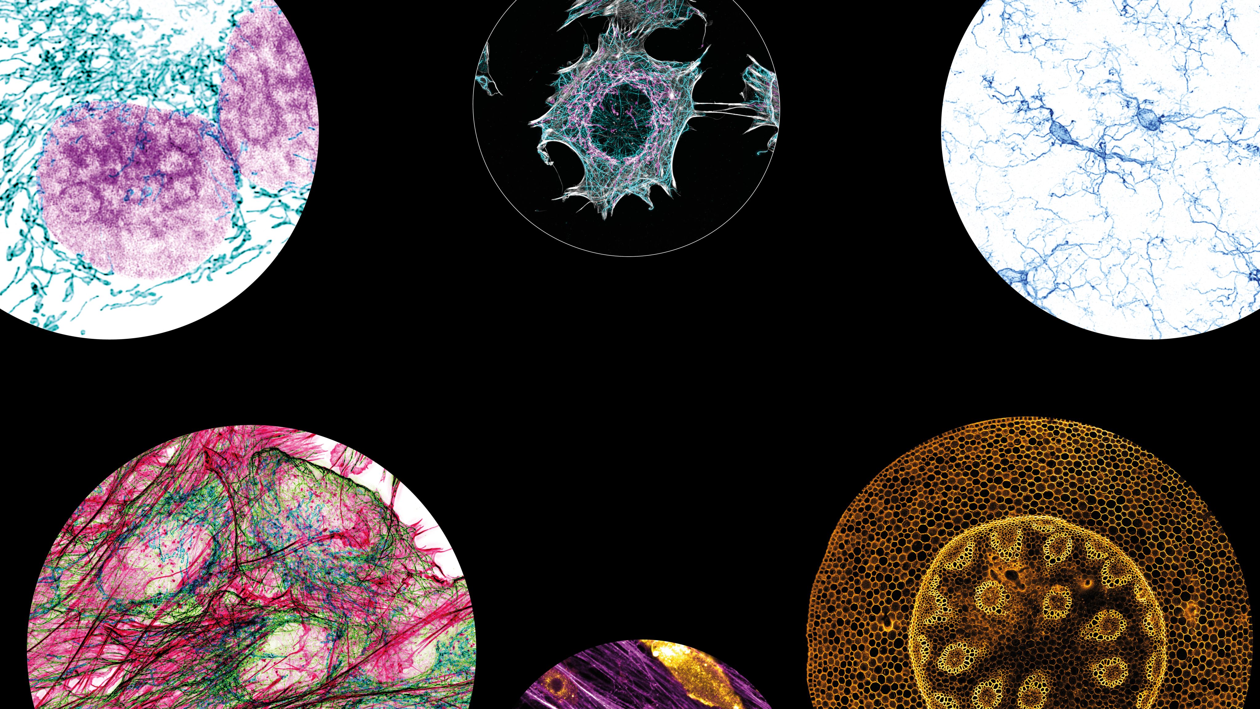
Microscopic imagery

Human fibroblast cells, stained for mitochondria (magenta), microtubules (cyan) and actin (grey), imaged by three dimensional structured illumination microscopy.

Cell migrating on extracellular matrix, grabbing at filaments, imaged on a confocal microscope.

Full cross-section of the stem of Convallaria majalis (Lily of the Valley), imaged on a confocal microscope.
Imperial is the magazine for the Imperial community. It delivers expert comment, insight and context from – and on – Imperial's engineers, mathematicians, scientists, medics, coders and leaders, as well as stories about student life and alumni experiences.
This story was published originally in Imperial 55/Winter 2023–24.